
Eighteen-Month Imaging after my Lumpectomy: Bilateral Mammogram with a Surprise Ultrasound
My eighteen-month imaging after my lumpectomy was scheduled to be a single mammogram on my right breast. This was the one that had DCIS in 2019. I ended up getting a bilateral mammogram with lots of images taken, followed by a series of ultrasounds. My anxiety ramped up during the imaging process, especially as the radiologist continued to order more views.
Was I going to get breast cancer, round two, for my summer vacation?
Thankfully, No. But there is much more to the story than that.
I’ll share my imaging story and also a few takeaways at the end that I hope will be encouraging if you are facing a medical procedure in the future.
Breast imaging has been an anxiety-producing process to go through after my lumpectomy in 2019. I am committed to getting the imaging done, but I feel scanxiety each time the appointments creep up. This was my third post-lumpectomy round of imaging, and it was the most challenging that I have gone through thus far.
Right Mammogram Turns into Bilateral Mammogram
“I’d like to do a bilateral mammogram on you since you are due for both sides, and I don’t want you to need to come back in again. Let’s do it now since you are here,” said the mammogram tech after she pulled up my chart in the imaging room.
My heart sank. I was hoping to have one mammogram on my right and didn’t think I would need a check-up on my left breast.
“Ok, but will it be a diagnostic mammogram?” I asked. With diagnostic imaging, I would have the ability to discuss things with the radiologist before I left the office. If it was a screening mammogram, I would receive the results in my online chart or the mail.
Thankfully, it would be a diagnostic mammogram, and I would get the results that day.
The tech got out the marking tape to place over my lumpectomy scars. This tape is a guide for the radiologist during the interpretation of the imaging. There were several rolls of marking tape for the tech to choose from on the wall of the imaging room, and I wasn’t sure what they were all for. She placed the one that was like a string with oval adhesives around it on me.
Side Note: If you are curious about why these types of markers are used during mammography and other information, check out Beekley Medical’s resource library. I also learned that they manufacture the MRI aromatherapy packets that the radiology team used during my breast MRIs.
The radiology tech took two images of each of my breasts. Then, she went to talk to the radiologist to see if he wanted additional views.
I sat down in the chair and pulled out my phone. I usually don’t bring it with me to the radiology room, but I had a feeling that I might be waiting a while in between scans. I was also feeling anxious. I resumed my game of Disney Emoji and waited for her to return. Playing games is one of the ways that I cope with my anxiety during medical appointments.
Follow-up Mammograms- Zooming into an Area on my Left
After a few minutes, the tech returned. The radiologist had requested additional imaging and different views for my breasts.
The tech changed out the mammography paddles for ones of a different size. One of the paddles she used was a small one, allowing the radiologist to better view a particular area. This paddle was not comfortable for me at all, but I knew that it was only going to be a few moments of compression.
I struggled with the discomfort during this round of mammography. At the end of May, I re-injured my left shoulder during my workouts. In 2017 I impinged my shoulder trying to carry a heavy suitcase up several flights of stairs in Paris. Working out too vigorously in the last few months had activated the injury again. I did not enjoy the arm stretching required to get the images on my left breast.
After this second round of mammography, the tech left the room again. She returned with the radiologist. He greeted me with a smile behind his mask and then went over to the imaging computer to discuss the additional imaging he wanted.
I wasn’t sure if he would say anything to me, but I had questions for him, so I spoke up.
“So, what is going on?” I asked once he was done with the conversation.
He told me that there was nothing of concern on my right- the side with the DCIS in 2019. However, he had noticed some density changes in my left breast. He wanted to obtain additional mammography images to compare with my previous imaging. He also wanted to add in an ultrasound as well.
“Can you do the ultrasound today?” I asked.
“Yes, we will do that as soon as we are done with the mammogram,” was his reply. I breathed a sigh of relief. At least I would have the answers before I left the building.
My “Interesting” Left Breast
In 2019, during my diagnosis process, a breast MRI revealed three findings that needed additional biopsies. These ultimately ended up being benign. But, the discovery and subsequent biopsies meant a delay in my surgery.
What was going on in my left breast, and was I going to need more biopsies?
Time for an Ultrasound
Once the tech finished up the mammograms, I left that room and went to the ultrasound room. I settled in on the table and tried to calm my nerves.
I had been texting Dave between the scans because I would be at the office for longer than I initially thought. I kept the messaging off of the family chat. I hoped this would all be nothing, and I didn’t want to worry the kids unnecessarily.
The tech began the scan on my left breast. I could see that she had my previous scans on another computer monitor and used her wand to get current images of the findings. She took several pictures and then told me she needed to check in with the radiologist.
I was once again left alone in the imaging room for a few minutes while they chatted. Out came my phone again, and I gave Dave the latest update. Then I played a terrible round of Emoji because by this time I was full of anxiety.
She entered again, took a few more images, and then went back out to chat.
The team certainly got points for being thorough during these scans.
The tech came back and said that they had all they needed and I could get dressed. The radiologist would come to talk to me once I was out of my gown.
She walked me back to the dressing rooms, and I closed the door to get dressed.
The Scanxiety was Awful
I could feel my heart pounding in my chest as I was getting my clothes on. Why wouldn’t the radiologist come in right after the scans and talk to me? What did they see? How many biopsies would I need?
I got dressed and sat down in the private waiting area. At this point, I wasn’t able to play any fun games on my phone because my body was tense, my heart was beating fast, my legs were shaking, and my thoughts were spinning. I decided to take a photo of the pretty bromeliad in the waiting area.

Results Time
“Jennifer,” called the radiology tech, “you can come back now.”
I headed back into the ultrasound room, and the radiologist entered—time for the results.
“It is all benign,” he said. “We noticed that there were density changes from the imaging in December, and we needed to check the findings with the previous imaging we had on your file.”
I breathed out a huge sigh of relief. I wasn’t getting breast cancer this summer.
The radiologist was able to use my previous imaging from fall 2019 and December 2020 and make comparisons. There were no significant changes to the findings. My breasts were denser during this imaging, which makes sense since I am no longer on the tamoxifen.
All of the biopsies and imaging done during my diagnosis process in 2019 had given the radiologists enough historical information to be confident in the results of this imaging session.
Before the radiologist left, I asked him what the Birads imaging score would be on this round of imaging. He told me it was a 2: benign. He recommended follow-up screening in a year or sooner if my surgeon requested.
YAY!!!
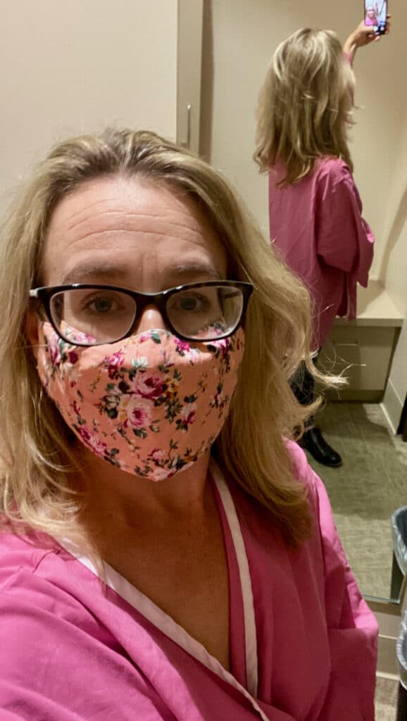
I skipped out of the office, headed to my car, pulled off my mask, sanitized my hands, and texted the family the good news.
I cried happy tears on the way home from the imaging center. I was so relieved.

Key Takeaways
My eighteen-month mammogram and ultrasound were challenging imaging sessions, and I wanted to share a few key takeaways after going through this. If you are facing a medical procedure and feeling anxious, I hope that these might be encouraging to you.
- Commit to the Process: This imaging was stressful, and I didn’t enjoy it. But I committed to following up regularly with my imaging when I chose a lumpectomy in 2019. Regular mammograms are a part of sticking to my commitment.
- Lots of Imaging Doesn’t Always Mean Cancer: The techs took many images of my breasts during this process. I was afraid that the number of images was an indication of new cancer, but it wasn’t.
- Scanxiety Sucks: I was nervous and anxious before, during, and immediately after the imaging. I didn’t enjoy it, and it wasn’t easy to manage.
- Bring your Phone: I don’t always bring my phone into the imaging room, but I’m so glad I did. I was able to pass the downtime with my games and stay in contact with my husband.
- Ask Questions: During diagnostic imaging, there is often an ability to discuss things with the radiologist. If you have questions, ask them. I’m glad that I took the time to ask which breast they were focusing on with the additional imaging. I was relieved it wasn’t the one that had already had DCIS.
- Plan Time to Decompress after the Appointment: I was spent emotionally after the imaging session. I needed to relax for a few hours after the imaging. I’m glad I planned and had nothing on my calendar after that appointment.
This imaging session was by far the most challenging round of follow-up imaging I have had to date. I’ve now been off of tamoxifen for seven months, and my breasts are back to their old level of density. I’m concerned about what the future will be in my left, but all I can do is continue to get imaging.
I will go back in December 2021 for more imaging, but until then, I will enjoy a lovely interval of time without worrying about biopsies or breast cancer.
Jennifer Douglas
Jennifer Douglas is an author, patient advocate, and DCIS breast cancer survivor. After navigating her own breast cancer journey in 2019, she began writing and encouraging others who were newly diagnosed. Her resources include her book, "A Breast Cancer Journey: Living It One Step at a Time," and her online support course, "Encourage: Breast Cancer and Beyond." Jennifer also actively supports patients through her online presence and direct involvement in communities and support groups, offering guidance and encouragement every step of the way.


5 Comments
Susan Budde
Looks like you had imaging every 6 months. My Mayo doctor said once a year and I was a stage 3A!!! I don’t agree with that but I have an appt with the Nurse Practioner tomorrow and I am going to request more frequent mammograms. They will be doing a contrast mammogram on my next one. I was diagnosed in 10/2020 and had a breast MRI in November but no scans since.
Jennifer Douglas
My surgeon likes imaging every 6 months for the first two years. This coming December will be 2 years, so we will see if he goes to once a year after that. The radiology department would be fine with once a year, but defers to my surgeon. I had an MRI and mammogram at 6 months, ultrasound and mammogram at 12 months and then the latest imaging with a mammogram and ultrasound. It is interesting how the different doctors want different frequencies. Keep me posted on your conversations with your team! Hope all goes well!
Ellen
I had a left lumpectomy in 1998 with radiation and chemo. Follow up imaging a year or so later showed an area of interest in my right breast. Eventually at 9 years out my annual mammogram was arranged as right side only, but the technician insisted on immediately getting doctor approval to do bilateral. (As long as you have a breast it should be checked, after all). She quite possibly saved me – left side had invasive DCIS, which otherwise would have been undetected at least another year. Left mastectomy done. It was 13 more years of mammography plus magnifying plus sonograms for that spot before the right side area of interest finally showed enough changes to treat with surgery. Now that I have had two mastectomies all that part is behind me.
Jennifer Douglas
So glad the tech insisted on doing the other side for you as well! Thank you for sharing your experiences- it highlights the importance for us all to keep getting our scans regularly!
Pingback: